December 15, 2020
Development of a Novel Tissue Clearing Reagent: The First Step Toward Application in Pathological Diagnosis
|
|
Associate Professor Shintaro Fumoto and his colleagues at the Nagasaki University Graduate School of Biomedical Sciences (Pharmaceutical Sciences) have developed a new tissue clearing reagent.
Key points
・Until now, tissue clearing methods with high transparency have generally used detergents to actively remove lipids, while tissue clearing methods that do not contain detergents have had insufficient transparency.
・In this study, we succeeded in development of a novel tissue clearing reagent that possesses both high transparency and preservation of lipid structures such as cell membranes. This reagent is a three-in-one method for achieving rapid (several hours) and efficient tissue clearing with a simple one-step clearing process.
・This reagent can also adjust pH in a wide range, making it possible to visualize the spatial distribution of low-molecular-weight fluorescent probes in tissues, which was previously difficult. It is expected to be applied to the analysis of spatial distribution of drug delivery systems such as liposomes in tissues and pathological diagnosis.
Details
Tissue is opaque because the refractive index is non-uniform due to the mixture of various components such as lipids, and light does not travel straight through it. Therefore, tissues are generally sliced thinly to observe the tissue structure. If the tissue can be made transparent, not only the tissue structure but also various phenomena such as the in vivo fate of antibody drugs, nucleic acids and gene medicines can be observed in 3D. Various tissue clearing technologies have been developed, but there is a kind of dilemma; methods with high transparency cannot retain lipid structures, while methods that preserve lipid structures have insufficient transparency.
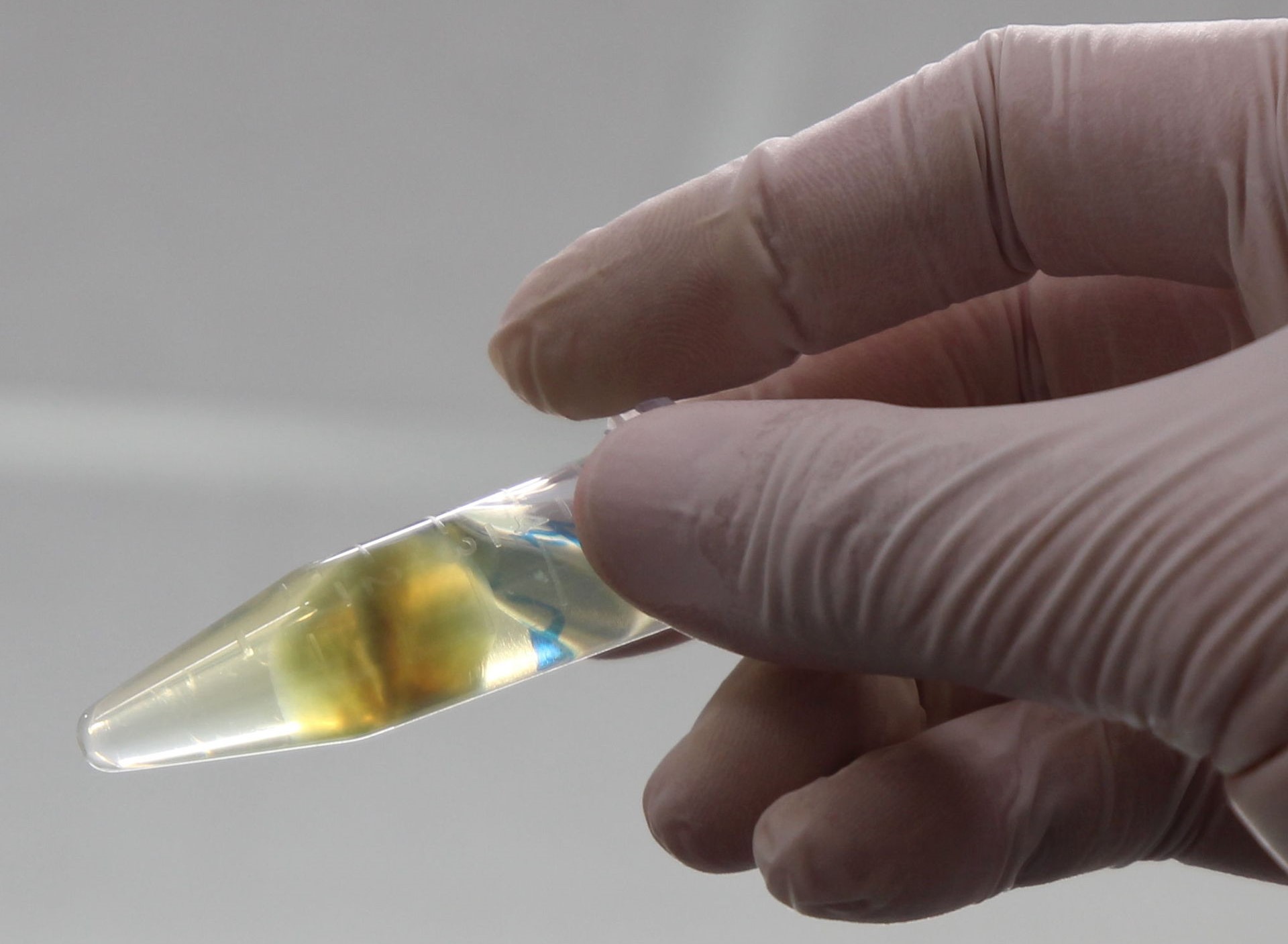
Therefore, Associate Professor Fumoto and his colleagues tried to develop an efficient tissue clearing reagent while maintaining the lipid structure. Taking a hint from the compounds used in conventional methods, they focused on polyethyleneimine, a polymer with amino groups that might have excellent tissue clearing efficiency. Although polyethyleneimine itself also has a tissue clearing effect, we achieved further tissue transparency by combining it with urea, which was used in conventional methods. We named it Seebest (SEE Biological Events and States in Tissues). Seebest does not contain detergents, can retain lipid structures including cell membranes, and boasts high transparency. In the past, it was common to immerse the cells in two or more different reagents, but with Seebest, only one reagent is needed, which is simple and time-saving. Since polyethyleneimine can buffer at a wide range of pH (5-11), which, combined with cell membrane retention, it allows various fluorescent probes to be retained in the cells for observation. Polyethyleneimine turns yellow in water over time, but we solved this problem by using propylene oxide-denatured polyethyleneimine.
In this study, we demonstrated the 3D-visualization of fluorescent proteins, vascular structures, liposomes and oxidative stress generation sites. In addition, it is expected to be applied to the kinetic analysis of fluorescently labeled antibody drugs in the body and, in the future, to the pathological diagnosis of biopsies in diseases such as non-alcoholic steatohepatitis (NASH) and cancer.
This study was conducted in collaboration with Professor Keisuke Ohta of Kurume University and Associate Professor Tasuku Hirayama of Gifu Pharmaceutical University. This study was published online in the international journal "Pharmaceutics" on November 9, 2020 (https://www.mdpi.com/1999-4923/12/11/1070)


